
Elastin Stain (Miller) An Extensive Educational Guide to Elastic Fiber Visualization in Histology and Tissue Research
Elastic fibers are fundamental structural components of many tissues, providing resilience, elasticity, and mechanical stability. Their proper organization and integrity are essential for the normal function of blood vessels, lungs, skin, and connective tissues. In laboratory research and histological studies, accurate visualization of elastic fibers is critical for understanding tissue architecture, remodeling processes, and experimental disease models.
Elastin Stain (Miller) is a classical and widely respected histological staining method designed to selectively demonstrate elastic fibers in tissue sections. This long-form educational article provides an in-depth overview of elastin biology, the scientific principles behind Miller’s elastin stain, and its broad applications in histology, pathology research, and biomedical investigation.
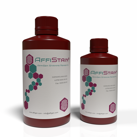
Elastic Fibers and Elastin: Structural Foundations of Tissue Elasticity
Elastic fibers are composite extracellular matrix structures composed primarily of elastin and microfibrillar proteins. They allow tissues to stretch and recoil repeatedly without losing structural integrity. Educational resources from the National Library of Medicine (https://www.ncbi.nlm.nih.gov) and the National Center for Biotechnology Information (https://www.ncbi.nlm.nih.gov/pmc) describe elastin as a highly durable protein that plays a key role in maintaining tissue flexibility and mechanical strength.
Academic overviews from Harvard Medical School (https://hms.harvard.edu) and Stanford School of Medicine (https://med.stanford.edu) emphasize that elastic fibers are particularly abundant in large arteries, pulmonary tissue, dermis, and ligamentous structures. Changes in elastin organization are frequently studied in experimental models of vascular remodeling, connective tissue biology, and aging research.
Why Stain for Elastin in Histological Research?
Histological staining remains one of the most powerful tools for visualizing tissue structure at the microscopic level. While routine stains such as hematoxylin and eosin provide general morphological information, they do not specifically highlight elastic fibers.
Educational materials from Yale School of Medicine (https://medicine.yale.edu) and Johns Hopkins University (https://www.hopkinsmedicine.org) explain that special stains for elastin are essential for:
-
Differentiating elastic fibers from collagen and other extracellular matrix components
-
Studying vascular wall architecture and integrity
-
Assessing connective tissue organization
-
Supporting morphometric and comparative tissue analyses
Elastin-specific staining techniques are therefore foundational tools in histology laboratories and research pathology workflows.
Overview of Elastin Stain (Miller)
Elastin Stain (Miller), also known as Miller’s elastic stain, is a classical histochemical method used to selectively stain elastic fibers in tissue sections. The stain produces strong contrast between elastic fibers and surrounding tissue elements, enabling clear visualization under light microscopy.
According to histotechnology references available through the National Institutes of Health (NIH) (https://www.nih.gov) and educational histology manuals hosted by University of Michigan Medical School (https://medicine.umich.edu), Miller’s method is valued for its reliability, reproducibility, and clarity of elastic fiber staining.
Scientific Principle of Miller’s Elastin Stain

The Elastin Stain (Miller) is based on the selective binding of dye complexes to elastin fibers within tissue sections. Elastin has unique chemical properties that allow it to retain certain dyes more strongly than surrounding connective tissue components.
Educational resources from Cold Spring Harbor Laboratory (https://cshl.edu) and MIT OpenCourseWare (https://ocw.mit.edu) explain that histochemical staining relies on differential chemical affinities between tissue components and staining reagents. In Miller’s method, elastic fibers are prominently stained, while collagen, muscle, and other structures provide contrasting backgrounds.
This selectivity allows researchers to clearly distinguish elastic fibers and analyze their distribution, density, and organization.
Typical Histological Workflow Using Elastin Stain (Miller)
A standard laboratory workflow for Miller’s elastin stain generally includes:
-
Tissue fixation – commonly formalin-fixed specimens
-
Embedding and sectioning – paraffin-embedded tissue sections
-
Deparaffinization and hydration
-
Application of Miller’s elastin staining reagents
-
Differentiation and counterstaining (if applicable)
-
Dehydration, clearing, and mounting
Best-practice histology workflows are consistent with recommendations published by the Centers for Disease Control and Prevention (CDC) (https://www.cdc.gov) and educational laboratory manuals from University of Washington Histology Core (https://depts.washington.edu).
Applications of Elastin Stain (Miller) in Research and Histology
Vascular Biology and Cardiovascular Research
Elastic fibers are critical components of arterial walls. Miller’s elastin stain is widely used to study vessel architecture, elastic lamina integrity, and structural remodeling in experimental vascular research. Educational content from the National Heart, Lung, and Blood Institute (NHLBI) (https://www.nhlbi.nih.gov) highlights the importance of elastin visualization in cardiovascular studies.
Pulmonary and Respiratory Tissue Studies
In lung research, elastin contributes to alveolar recoil and airway stability. Academic pulmonary research programs at University of California, San Francisco (UCSF) (https://pulmonary.ucsf.edu) describe elastin staining as an essential technique for examining lung tissue architecture in experimental models.
Connective Tissue and Skin Research
Elastic fibers are abundant in dermal tissue and connective structures. Educational dermatopathology resources from University of Oxford (https://www.ox.ac.uk) and University of Cambridge (https://www.cam.ac.uk) emphasize elastin stains for studying skin elasticity, connective tissue organization, and extracellular matrix composition.
Comparative Histology and Teaching Laboratories
Miller’s elastin stain is frequently used in academic teaching laboratories to demonstrate extracellular matrix components. Histology curricula from Princeton University (https://molbio.princeton.edu) and University of Michigan (https://research.umich.edu) incorporate elastin staining to help students understand tissue mechanics and structure–function relationships.

Advantages of Elastin Stain (Miller)
Elastin Stain (Miller) offers several advantages for laboratory and educational use:
-
High contrast visualization of elastic fibers
-
Clear differentiation from collagen and smooth muscle
-
Compatibility with routine paraffin sections
-
Reproducible and well-established methodology
These strengths are highlighted in histological technique guides supported by the National Academies of Sciences (https://www.nationalacademies.org).
Interpretation of Stained Sections
When properly performed, Miller’s elastin stain produces intensely stained elastic fibers that appear dark against a lighter background. Researchers can assess:
-
Fiber continuity and fragmentation
-
Density and spatial distribution
-
Structural organization within tissues
Guidance on histological interpretation and quality control is provided by the NIH Office of Research Integrity (https://ori.hhs.gov) and educational pathology resources from National Cancer Institute (NCI) (https://www.cancer.gov).
Conclusion
Elastin Stain (Miller) is a cornerstone histological technique for the selective visualization of elastic fibers in tissue sections. Its clarity, reliability, and strong educational foundation make it an indispensable tool in histology laboratories, pathology research, and academic teaching environments.
By enabling precise examination of elastic fiber organization, Miller’s elastin stain supports advanced research in vascular biology, pulmonary studies, connective tissue research, and comparative histology. Its long-standing use in educational and government-supported research institutions reinforces its value as a trusted and authoritative staining method for high-quality scientific investigation and educational blogging.
Educational and Governmental Resources Referenced
This article integrates authoritative educational and governmental sources, including:
Recent Posts
- Competitive Inhibition ELISA Kits: Principles, Applications, and Advantages in Immunoassay Research
- APOE ELISA Kits: Sensitive Immunoassays for Apolipoprotein E Quantification in Molecular Biology Research
- Amyloid ELISA Kits: Sensitive Immunoassays for Amyloid Protein Quantification in Molecular Biology Research
- AAV ELISA Kits: Reliable Quantification Tools for Adeno-Associated Virus Research
- Advanced Tools for Oxidative DNA Damage Research and Molecular Biology Studies
Categories
| M | T | W | T | F | S | S |
|---|---|---|---|---|---|---|
| 1 | 2 | 3 | 4 | 5 | 6 | |
| 7 | 8 | 9 | 10 | 11 | 12 | 13 |
| 14 | 15 | 16 | 17 | 18 | 19 | 20 |
| 21 | 22 | 23 | 24 | 25 | 26 | 27 |
| 28 | 29 | 30 | ||||